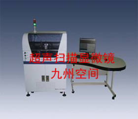
The structural morphology of high molecular polymers is divided into microscopic structure and macroscopic structure. The microstructure morphology refers to the aggregation state of the polymer on the microscopic scale, such as the crystalline state, the liquid crystal state or the disordered state (liquid state), and the uniformity of crystal size and nano-scale phase dispersion. The microstructure state of the polymer determines its macroscopic mechanical and physical properties, and further defines its application and scope. The macroscopic structure refers to the morphology of the surface of the polymer, the shape of the section, and the distribution of the micropores (defects) contained on the macroscopic or submicroscopic scale. Observing the surface structure, the cross section and the internal microphase separation structure of the solid polymer, the distribution of micropores and imperfections, the crystal size, traits and distribution, and the uniformity of the nanometer phase dispersion, etc., will improve the processing of the polymer. The preparation conditions, the choice of blending components, and the optimization of material properties provide data.
The characterization methods and equipment for the morphology of polymer polymers include:
1. Polarized light microscope (PLM)
By utilizing the optical properties of the polymer liquid crystal material, different polymer liquid crystals can be observed by a polarizing microscope, and the type of the polymer liquid crystal is qualitatively determined by the texture image of the liquid crystal.
2. Metallographic microscope
The metallographic microscope can observe the submicroscopic structure of the surface of the polymer and determine the defects and macro defects in the polymer. Stereoscopic optical microscopy is usually used to observe the structural features of the surface and section of a polymer polymer body, and provides important information for optimizing the production process and performing damage failure analysis.
3, stereo microscope
When using a stereo microscope, care should be taken not to introduce further damage into the sample being observed during sampling. When using a metallographic microscope, the sample to be tested needs to be first fixed in a mold and then cast into a cylindrical sample with a resin. The ground of the cylinder is the surface to be measured. The surface to be tested is polished and polished into a mirror surface and placed on a metallographic microscope. The clarity of the submicroscopic morphology of a polymer depends on the quality of the surface to be polished.
4. X-ray diffraction
The crystal structure and liquid crystal structure information of the polymer can be obtained by wide-angle or small-angle diffraction of X-rays. For details, see the crystalline state of the polymer and the liquid crystal state of the polymer.
5. Scanning electron microscopy (SEM)
Scanning electron microscopy uses an electron beam to scan a polymer surface or section to produce an image of the surface of the object to be measured on a cathode ray tube (CRT). For conductive samples, it can be adhered to the copper or aluminum sample holder with conductive adhesive to directly observe the measured surface; for the insulating sample, a conductive layer (gold, silver or carbon) needs to be sprayed on the surface in advance.
The surface morphology of the polymer can be observed by SEM; the phase separation size and phase separation pattern shape of the surface of the polymer multiphase system filling system; the fracture characteristics of the polymer section; the size and uniformity of the nanometer-scale dispersed phase in the nanomaterial section; .
6. Transmission electron microscopy (TEM)
Transmission electron microscopy can be used to characterize the morphology of the internal structure of the polymer. The polymer sample to be tested is uniformly dispersed on the surface of the sample support film by a suspension method, a spray method, an ultrasonic dispersion method, etc., or a solid sample of the high molecular polymer is cut into a thin 50 nm by an ultramicrotome. Sample. The prepared sample was placed on a sample holder of a transmission electron microscope, and the structure of the sample was observed by TEM. The crystal structure, shape, and distribution of the crystal phase of the polymer can be observed by TEM. High-resolution transmission electron microscopy can observe crystal defects of high molecular polymer crystals.
7. Atomic Force Microscopy (AFM)
Atomic force microscopy uses a microprobe to scan the surface of the polymer being tested. When the probe tip approaches the sample, the probe tip is deformed by the van der Waals force of the sample molecule. Due to the different molecular types and structures, the van der Waals forces are also different in size, and the deformation of the probes in different parts also changes, thereby "observing" the morphology of the polymer surface. Since the scanning of the polymer surface by the atomic force microscope probe is three-dimensional scanning, the three-dimensional morphology of the surface of the polymer can be obtained.
Atomic force microscopy can observe the morphology of the polymer surface, the conformation of the polymer chain, the order and orientation of the polymer chain, the size and uniformity of the phase separation size in the nanostructure, the crystal structure, shape, crystal formation process, etc. information.
8. Scanning tunneling microscope (STM)
Similar to atomic force microscopy, scanning tunneling microscopy also scans the surface of the conductive polymer to be tested with tiny probes. When the probe and the conductive polymer are close to each other, under the action of an external electric field, they will be in the conductive polymer and probe. Between, a weak "tunnel current" is generated. Therefore, the distribution of the occurrence of the "tunnel current" on the surface of the polymer can be measured, and the topographical information of the surface of the conductive polymer can be "observed".
The scanning tunneling microscope can obtain the surface morphology of the polymer, the conformation of the polymer chain, the order and orientation of the polymer chain, the size and uniformity of the phase separation size in the nanostructure, the crystal structure and shape. Compared to atomic force microscopy, scanning tunneling microscopes can only be used for the observation of conductive polymer surfaces.

A wrecker tow truck, rotator wrecker is used to pull broken or damaged cars and vehicles to the repair shop, impound lots. we have a wide selection of tow truck, wreckers, rollbacks, rotators and heavy duty tow truck for sale, short lead time, help you start a tow truck business in shortest time. as a leading tow truck manufacturer, we have have a huge selection of tow truck parts and accessories available in the meantime.
Choose a good quality wrecker tow truck, recovery truck could help you better start your business for long run.
Wrecker Tow Truck,Recovery Tow Truck,Iveco Tow Truck,Iveco Recovery Truck
Hubei Jiangnan Special Automobile Co., Ltd , https://www.dingshunglass.com